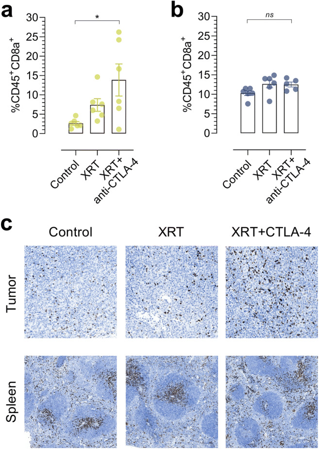
Fig. 4.

Flow cytometric analysis of a tumors and b spleens harvested from control, XRT-, and XRT + anti-CTLA-4-treated mice on day 8 following treatment initiation (N = 6/group for tumor, N = 5–6/group for spleen). c Representative immunohistochemical staining of CD8a+ in paraffin-embedded tumors and spleens harvested from control, XRT-, and XRT + anti-CTLA-4-treated mice on day 8 following treatment initiation. Data are presented as mean ± SEM. The significance level is indicated by asterisks (*). *p < 0.05, **p < 0.01, ***p < 0.001, ****p < 0.0001. XRT, external radiation therapy.
