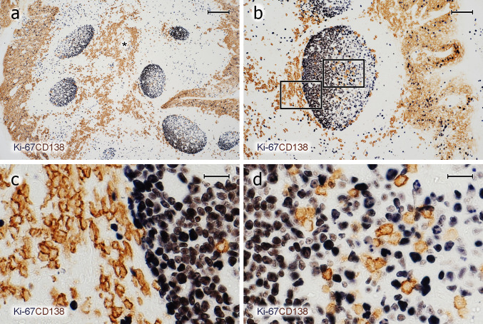
Fig. 3.
PCs inside and outside GCs do not proliferate. a–d Visualisation of Ki-67 (mAb MIB-5, blue-black) followed by staining for CD138 (mAb BB-4, brown) shows that the vast majority of PCs inside and outside GCs is Ki-67−. c, d Magnifications of the areas indicated in b. The entire crypt epithelium is also CD138+ (a, b). It covers both sides of the specimen in a and the right side in b. The brown cells in the central part of a represent PCs accompanying the central connective tissue septum (asterisk) between two crypts. a–d 21-year-old female. Scale bars 200 µm (a), 100 µm (b), and 20 µm (c, d)

