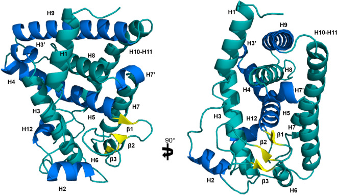
Figure 3.
Structure of DimDAF-12 ligand binding domain model. The model of DimDAF-12_LBD (A and B) was constructed using Modeller and the coordinates of the crystal from Ancylostoma ceylanicum DAF-12 (pdb:3up0) and Strongyloides stercoralis DAF-12 (3gyt). Similar to the other members of the nuclear receptor family, DimDAF-12 exhibits 13 helices and three β-strands, which are packed in a three-layer α-helical sandwich to create a ligand binding pocket, where the DAs were found to bind in the case of AceDAF-12 and SstDAF-12. α-helices were either shown in teal or marine to distinguish them, β-strands were colored in yellow and the loops in teal. All images were generated using PyMol 1.3 (https://pymol.org/2/).

