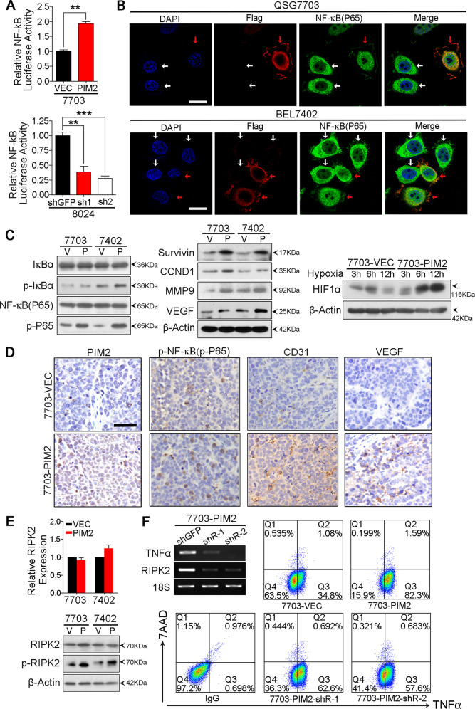
Fig. 5. PIM2 overexpression activated NF-κB signaling pathway.
a Relative NF-κB signaling pathway activity of empty vector-transfected and PIM2-transfected QSG7703 cells, and relative NF-κB signaling pathway activity of shGFP-transfected and shPIM2-transfected PLC8024 cells detected by luciferase reporter assay. The result is shown as mean ± SD of three independent experiments. **p < 0.01, ***p < 0.001, independent Student’s t test. b Immunofluorescence images of QSG7703 and BEL7402 cells transfected with Flag tagged PIM2 (indicated by red arrows) or their parental cells (indicated by white arrows). Cells were double stained with Flag antibody (red) and NF-κB (P65) antibody (green). Nuclei were labeled with DAPI (blue). Scale bar stands for 20 µm. c Expression of NF-κB (P65), p-NF-κB (p-P65), IκBα, p-IκBα, and NF-κB signaling pathway target genes, Survivin, CCND1, MMP9, VEGF, and HIF1α detected by Western Blot. β-Actin served as an internal control. V represents empty vector-transfected group and P represents PIM2-transfected group. d IHC images of paraffin sections from xenografts formed by 7703-VEC and 7703-PIM2 injection. Scale bar stands for 20 µm. e Relative expression level of RIPK2 in empty vector-transduced and PIM2-transduced 7703 and 7402 cells detected by qRT-PCR, and expression of RIPK2 and p-RIPK2 in empty vector-transduced and PIM2-transduced HCC cells detected by Western Blot. V represents empty vector-transduced group and P represents PIM2-transduced group. f Expression of RIPK2 and TNFα in shGFP-transduced and shRIPK2-transduced 7703-PIM2 cells detected by semiquantitative RT-PCR,18S rRNA was used as an internal control; and expression of TNFα in 7703-VEC, 7703-PIM2, 7703-PIM2-shRIPK2-1, and 7703-PIM2-shRIPK2-2 detected by flow cytometry.

