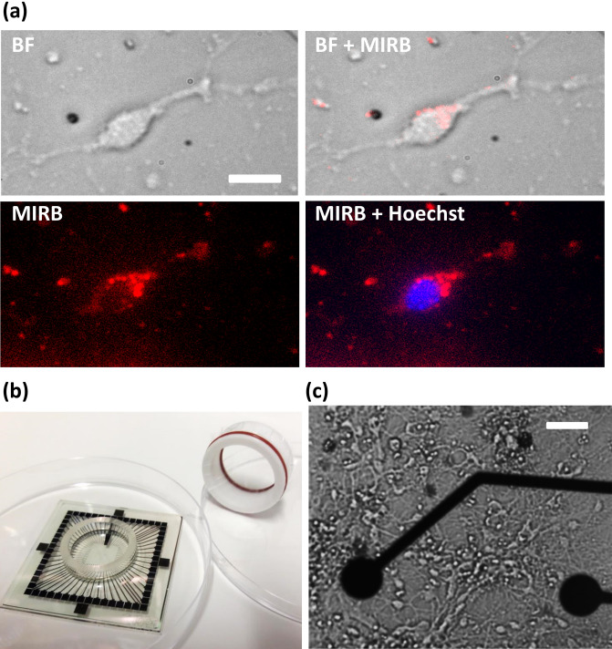
Figure 1.
MIRB uptake by murine neurons in an MEA culture chamber. (a) Localization of MIRB nanoparticles in neurons. DIV19 neurons were incubated for 2 h with 10 µg/ml MIRB nanoparticles before the particles were washed out. Both bright field and fluorescence images were acquired at 24 h subsequent to the start of this incubation (DIV20). The fluorescence image of MIRB nanoparticles is shown in red; the fluorescence image of Hoechst 33342 stain is shown in blue. Scale bar is 20 µm. (b) A photograph of a microelectrode array (MEA) and specialized MEA lid used in this study. The MEA is placed inside a 100 mm culture dish for size reference. (c) A bright field image of a typical neuron culture (DIV19) plated in a MEA chamber. Scale bar is 30 µm.

