
Figure 7.
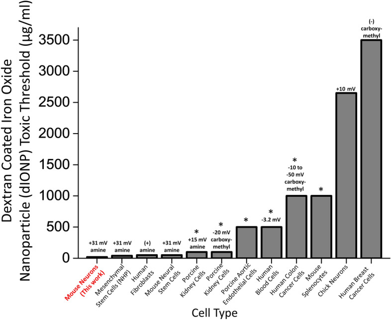
Dextran-coated iron oxide nanoparticle toxicity threshold for different cell types, zeta potentials, and functionalization groups. A comparison of threshold doses generating significant toxicity after exposure to ~ 10 nm-sized dextran coated iron oxide nanoparticle (dIONP). Each column lists the organism, tissue, particle functionalization and zeta potential: mouse neurons (this work) (amine functionalization), nonhuman primate mesenchymal stem cells32 (amine functionalization), human fibroblasts61 (amine functionalization), mouse neural stem cells15 (amine functionalization), porcine kidney cells62 (amine and carboxymethyl functionalization separately tested), porcine aortic endothelial cells38 (dextran only), human erythrocytes, monocytes, and leukocytes26 (dextran only), human colon cancer cells63 (carboxymethyl functionalization), mouse splenocytes60 (dextran only), primary chick neurons52 (dextran only), and human breast cancer cells64 (carboxymethyl functionalization), A (+) or (−) not accompanied by a numerical value indicates that the zeta potential was not reported in a given column’s study, but that we interpreted the amine and carboxymethyl functionalization as having a positive and negative charge respectively (as is standard). The absence of a zeta potential value indicates that the study used a dextran coating without functionalization, so that the sign of the surface charge is ambiguous. A “*” above a column indicates the highest dIONP dose value tested in that study, but that no gross toxicity effects were observed even at this dose. See each column’s reference for more information on the specific toxicity assays used for each study.