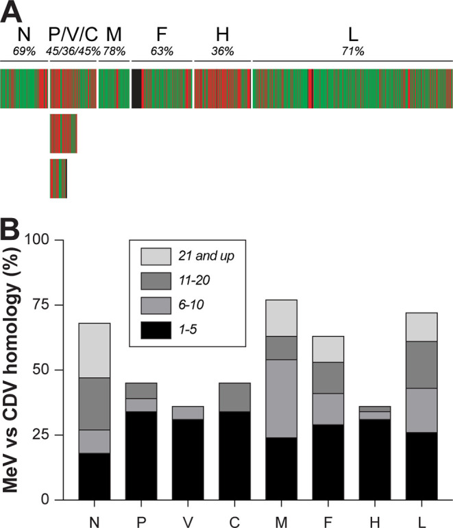
FIG 1.

Conservation between MeV and CDV. (A) Heat map indicating homology between MeV and CDV for the respective open reading frames (ORFs). Green represents fully homologous residues, and red shows nonhomologous residues. Proteins and total percentage of conservation are indicated above the heat map. (B) Stretches of fully conserved homologous amino acids when comparing all ORFs from MeV and CDV, containing potential cross-reactive T cell epitopes. Stretches of 1 to 5, 6 to 10, 11 to 20, and 21 or more homologous amino acids are indicated in shades of gray. MeV, measles virus; CDV, canine distemper virus; N, nucleoprotein; P, phosphoprotein; M, matrix protein; F, fusion protein; H, hemagglutinin; L, large protein or polymerase.
