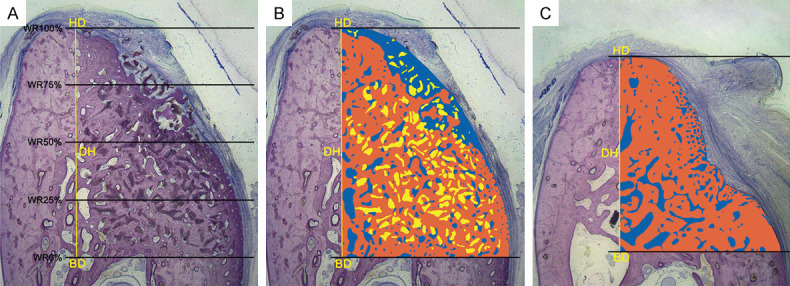
Figure 3.

Methods of histomorphometric analysis. A. The following landmarks were identified in the stained sections: the bottom of the defect (BD) and the highest point of the alveolar ridge of the defect (HD). The defect height (DH, mm) was measured from the HD to the BD, and the width of the newly formed ridge (WR, mm) was measured at 0%, 25%, 50%, 75%, and 100% of the DH. B and C. The colored area, measured from the BD to the HD, represents the region of interest (ROI) of the augmentation procedure. Within the ROI, the different colors represent different structures: Orange = new bone; blue = non-mineralized structures; and yellow = residual material. The area (mm2) and proportion (%) of the different structures were calculated (McNeal stained, original magnification 10×).
