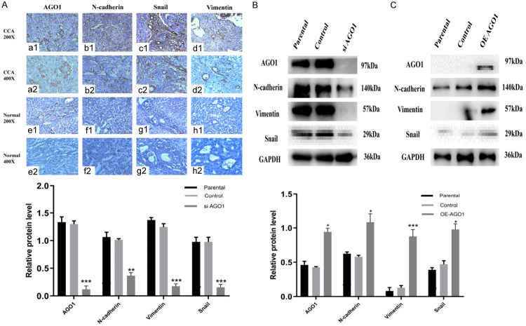
Figure 3.
Immunohistochemical staining and Western blotting for AGO1, N-cadherin, Snail and vimentin. We examined the expression of EMT-related proteins in tissues and cell lines. The expression of AGO1 is positively correlated with the expression of EMT-related proteins. A. a1, a2: Positive AGO1 expression in CCA tissue. e1, e2: Negative AGO1 expression in adjacent tissue. c1, c2: Positive N-cadherin expression in CCA tissue. f1, f2: Negative N-cadherin expression in adjacent tissue. c1, c2: Positive Snail expression in CCA tissue. g1, g2: Negative Snail expression in adjacent tissue. d1, d2: Positive Vimentin expression in CCA tissue. h1, h2: Negative Vimentin expression in adjacent tissue. (magnification: ×200, ×400). B. Western blot analysis of EMT signaling molecules (N-cadherin, Snail and Vimentin) in AGO1 silenced Hucct1 cell line. C. Western blot analysis of EMT signaling molecules (N-cadherin, Snail and Vimentin) in AGO1 overexpressed QBC939 cell line. Representative of three independent experiments.

