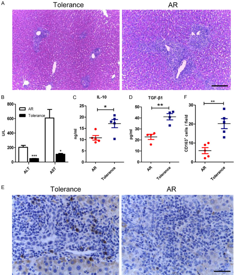
Figure 1.

The infiltration of the CD163-positive cells increased in the tolerant liver grafts. A. Representative images of HE staining of liver allografts in the tolerance and the AR group 7 days following transplantation with original magnifications of ×100. Characteristics like portal inflammatory cell infiltration, bile duct damage and endothelial inflammation significantly reduced in the tolerance group. Scale bar in right lower corner represents 100 µm. B. Liver functions were assessed on day 7 after transplantation. Both ALT and AST were significantly lowered in tolerance group. C, D. ELISA was used to detect serum IL-10 and TGF-β1 levels of the recipients in both groups. Both anti-inflammatory cytokines were significantly increased in the tolerant recipients. E. Illustrating IHC microscopic finding for identification of CD163 positive cells (brown color) with original magnifications of ×400. Scale bar in right lower corner represents 25 µm. F. Analytical results of the numbers of CD163 positive cells. The numbers of CD163 positive cells in the AR group were less than that in the tolerance group. All statistical analyses were performed by an unpaired t-test. Data are presented as the mean ± SD. (n = 5, *P < 0.05, **P < 0.01, ***P < 0.001).
