Figure 2.
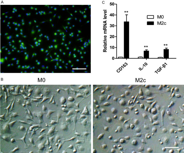
M2c BMDMs were successfully induced in vitro. A. The bone marrow derived cells were examined by immunofluorescence staining with anti-CD68 antibody after being stimulated by MCSF for 7 days. Nearly all cells expressed CD68, the specific rat macrophage marker (Magnification, 200). Scale bars in right lower corner represents 50 µm. These cells were then stimulated by PBS or dexamethasone for 24 h for M0 or M2c polarization. B. Representative images of the M0 and the M2c with original magnifications of ×400. Scale bar in right lower corner represents 25 µm. C. M2c polarization markers expression determined by qRT-PCR. The expression levels of CD163, IL-10, TGF-β1 in the M2c macrophages were significantly higher than those of the M0 macrophages. The statistical analyses were performed by an unpaired t-test. Data are presented as the mean ± SD. (n = 3, **P < 0.01).

