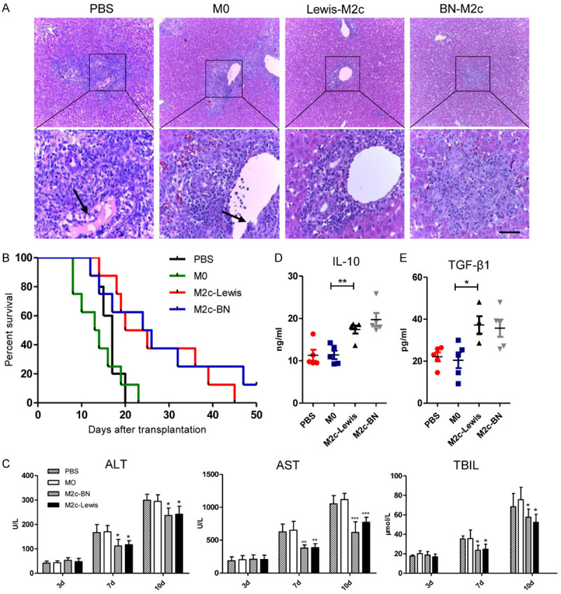
Figure 3.

Infusion of the M2c BMDMs alleviated acute rejection in rat liver transplantation. The M0, Lewis-M2c, BN-M2c macrophages (3×10⁶ cells per rat) or PBS were infused into recipients through portal vain immediately following OLT. A. Representative images of HE staining of liver allografts 7 days after liver transplantation with original magnifications of ×100 (upper panel) and ×400 (lower panel). The lymphocytes infiltration, bile duct damage and endothelial inflammation significantly reduced in recipients received the M2c macrophages, while no significant differences were observed between the M0 group and the PBS group. The black arrows show endothelial inflammation. Scale bar in right lower corner represents 25 µm. B. The survival analysis of the recipients was monitored and performed by the Breslow-Gehan-Wilcoxon test. The survival time of the recipients was significantly prolonged in the M2c infusion groups compared with that in the M0 and the PBS infusion group. C. Liver functions on day 3, 7, and 10 following liver transplantation. In comparison with the M0 and the PBS infusion group, ALT, AST, and TBIL in the M2c infusion groups were significantly improved. D, E. Serum IL-10 and TGF-β1 levels were quantified by ELISA and were significantly reduced in the M2c infusion groups compared with those in the M0 and the PBS infusion groups. No significant differences were observed between the M0 and the PBS infusion groups. All statistical analyses except survival rate were performed by one-way ANOVA, followed by post hoc test of Tukey’s (n = 5 for each group). Data are presented as the mean ± SD. *P < 0.05, **P < 0.01, ***P < 0.001 vs M0 group.
