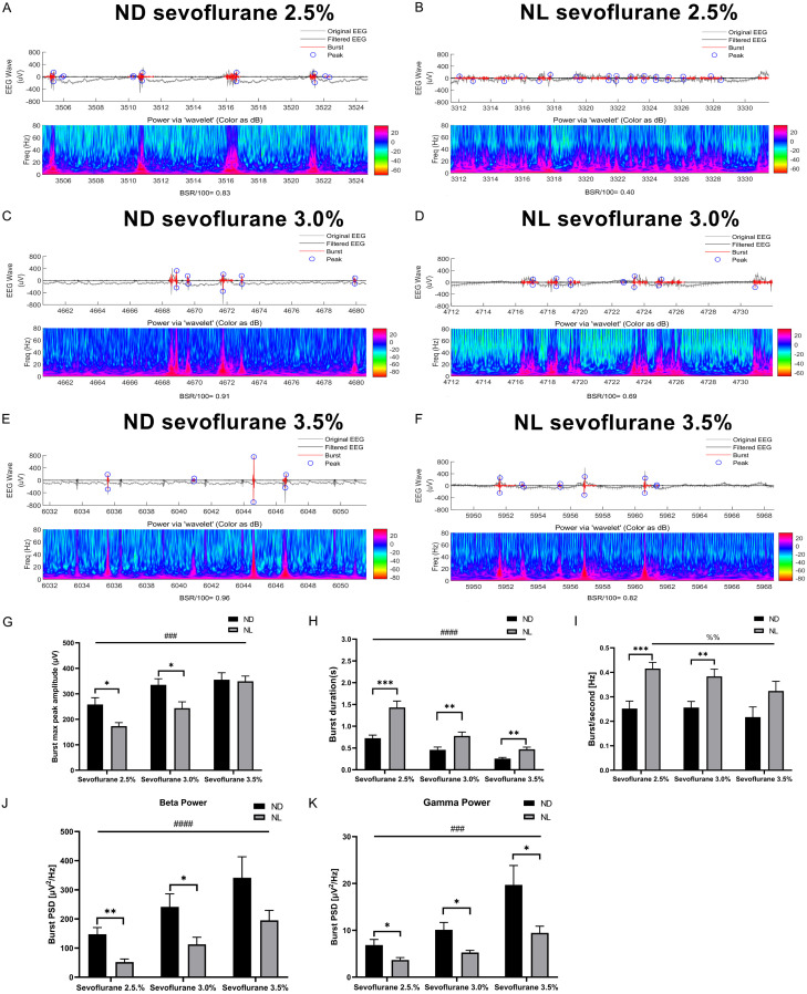
Figure 5.
Effect of acute continuous nocturnal light exposure (ACNLE) on spontaneous bursts under sevoflurane anesthesia. (A-F), representative EEG traces (up) and burst power (down) at increasing concentrations of sevoflurane for groups ND and NL. When the anesthetic concentration is increased, a clear burst-suppression pattern is induced. In the EEG trace, both the original EEG (gray line) and the filtered EEG (black line) are presented. Black on the filtered EEG trace indicates suppression, and red indicates a burst. Blue circles indicate the maximum and minimum amplitude values within each individual burst. (G-K), quantitative burst analysis obtained on identified burst episodes during burst suppression to illustrate the effect of acute nighttime light exposure on burst maximum peak-to-peak amplitude (G); burst duration (H); burst frequency (I); and burst power in beta (J) and gamma (K) bands. The burst duration and burst frequency decreased with increasing sevoflurane concentration in both groups. The peak-to-peak amplitude and burst power in the beta (15-25 Hz) and gamma (25-80 Hz) bands were increased with increasing sevoflurane concentration in both groups. Compared to group ND, the analysis of individual bursts showed that burst duration and burst frequency were significantly increased in group NL. The burst maximum peak-to-peak amplitude and burst power in the beta (15-25 Hz) and gamma (25-80 Hz) bands were decreased in group NL (##P < 0.01, ###P < 0.001, ####P < 0.0001, main effect of sevoflurane concentration by two-way rANOVA; %%P < 0.01 main effect of sevoflurane concentration by one-way rANOVA; *P < 0.05, **P < 0.01, ***P < 0.001 compared with ND, n = 10 in each group, two-way rANOVA followed by Fisher’s LSD test). Sevo = sevoflurane. ND, nighttime administration of sevoflurane (20:00-24:00) in the dark; NL, nighttime administration of sevoflurane (20:00-24:00) with exposure to light.

