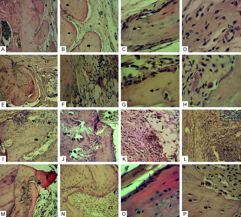
Figure 4.

(A-D) G0 Group: (A) Edge of defect exhibiting discreet projection-like bone ► formations and medullary spaces (ms). (B) Bone formation with osteocytes ➨ in their gaps and adjacent area filled with collagenized fibrous connective tissue (ct). (C) Bone trabeculae displaying osteoblasts → superficially and osteocytes ➨ within. (D) Collagenized fibrous connective tissue from the defect area, displaying numerous large fibroblasts ⇥, lymphocyte ⇢ and BMDblood vessel (bv). (E-H) G1 Group: (E) Bone projections ► formed on margin of defect. Connective tissue in defect area (ct) showing remnant intertwining of implanted biomaterial (b). (F) Area showing remnant of implanted biomaterial (b) where multinucleated giant cell ⇉ can be visualized in contact with the periphery of the biomaterial. (G) Bone trabeculae surface showing osteoblastic → paving and osteocytes ➨ inside. (H) Another field in a defect region showing osteoclast ►► on bone trabeculae surface and scarce mononuclear inflammatory cells *, lymphocytes ⇢, macrophages ⇟. (I-L) G2 Group: (I) Margin of defect showing bone fragments ► with irregular margins containing osteocytes ➨. (J) Field showing bone trabecula resorbed by numerous osteoclasts ►►. In detail, osteoclasts on bone surface. (K) Numerous multinucleated giant cells ⇉ in proximity to the implanted biomaterial. (L) Defect area showing intense inflammatory infiltrate *** permeating collagenized fibrous connective tissue (ct). In detail numerous mononuclear inflammatory cells are observed. (M-P) G3 Group: (M) Bone projections ► at the interface of the remnant bone edge and defect displaying osteocytes ➨ inside, scarce scattered inflammatory cells *. (N) Bone fragment ► surrounded by collagenized and well-cellularized fibrous connective tissue (ct). (O) Bone fragment presenting osteoblasts → in the periphery and osteocytes ➨ in the interior. (P) In the field near the edge of the defect, osteoclasts ►► are sometimes housed in gaps promoting bone resorption, lymphocytes ⇢, macrophages ⇟. G0 Group: Positive control critical defect (D) filled with blood clot; G1 Group: Critical defect filled with Hydroxyapatite (H) scaffold; G2 Group: Critical defect filled with zirconia (YSZ) scaffold; G3 Group: Critical defect filled with zirconia/hydroxyapatite (80/20) scaffold. (A, E, I-M) (HE 100 ×); (B, F, N) (HE 200 ×). (C, D, G, H, O, P) details (HE 400 ×).
