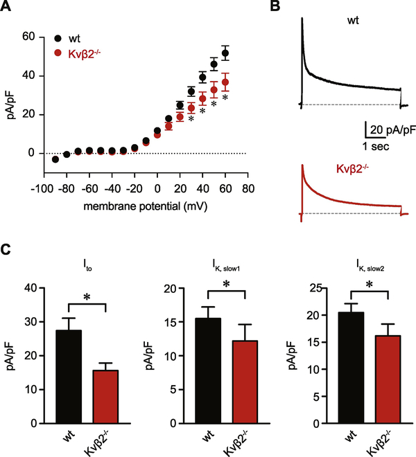
Figure 3: Suppressed Kv current density in cardiac myocytes from Kvβ2−/− mice.
(A) Summary current-voltage relationship obtained from cardiac myocytes from wild type and Kvβ2−/− animals. (B) Representative outward K+ current recordings normalized to cell capacitance (pA/pF) in response to a pulse depolarization to +50 mV from a holding potential of −80 mV in ventricular myocytes from wt and Kvβ2−/− animals. (C) Summarized current density magnitudes for Ito, IK,slow1, and IK,slow2 components, obtained from tri-exponential fitting of +50 mV-elicited current recordings, as shown in panel A, for wt and Kvβ2−/− myocytes (n = 15–19 cells from 4–6 mice). *P<0.05.

