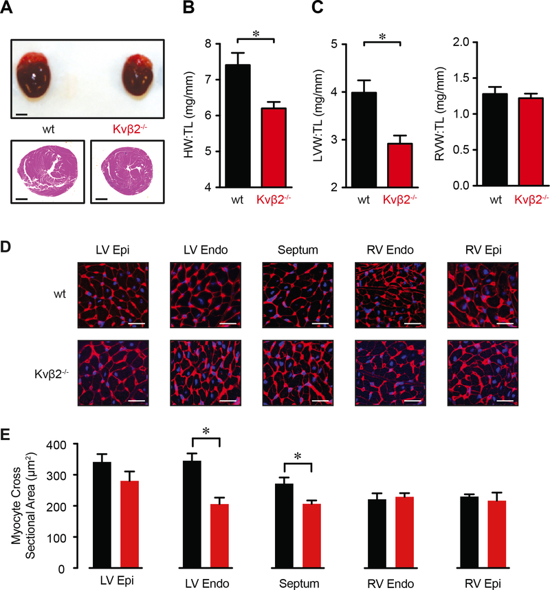
Figure 5: Loss of Kvβ2 alters heart morphology.
(A) Images showing whole hearts (top; scale bar = 2 mm) and hematoxylin and eosin stained transverse heart sections (bottom; scale bar = 1 mm). (B, C) Total heart weight (HW; B) and left and right ventricular weight (LVW, RVW; C) normalized to tibia length (TL) for hearts from wt (n = 12) and Kvβ2−/− (n = 13) animals. *P<0.05. (D) Representative images of WGA (membrane; red) and DAPI (nuclear; blue) staining in left and right ventricular (epi: epicardial; endo: endocardial) and septal regions in transverse mid-ventricular heart sections from wild type and Kvβ2−/− animals. Scale bars = 25 μm. (E) and summary of cardiomyocyte cross sectional area in regions as indicated in hearts from wild type and Kvβ2−/− animals. *P<0.05.

