FIGURE 6.
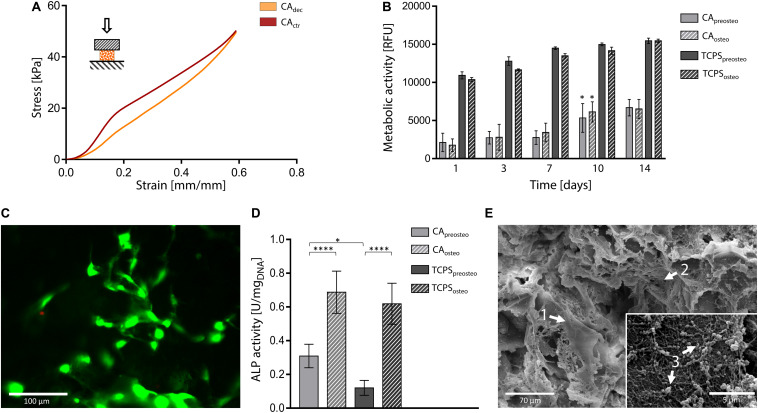
Decellularized carrot for bone tissue engineering. (A) Representative compression mechanical curve of carrot-derived scaffolds and control samples up to 60% strain. (B) Metabolic activity of MC3T3-E1 pre-osteoblast cultured up to 14 days on carrot scaffolds and TCPS plates. Cells were either induced toward osteogenic differentiation (i.e., CAosteo and TCPSosteo) or cultured in pre-osteogenic medium (i.e., CApreosteo and TCPSpreosteo). n = 4 per time point, one-way ANOVA: *p < 0.05 comparing the metabolic activity of the same sample at one time point to the previous time point. (C) Representative LIVE/DEAD staining of MC3T3-E1 cells cultured on carrot-derived scaffolds (scale bar: 100 μm). (D) ALP activity of MC3T3-E1 cells culture on decellularized carrots and TCPS for 14 days (values are normalized over DNA content); n = 4 per time point, one-way ANOVA: *p < 0.05, ****p < 0.0001. (E) SEM micrographs of osteogenic-differentiated MC3T3-E1 cells (arrow 1) cultured for 14 days on carrot-derived scaffolds. Nano-fiber nets (arrow 2), attributable to ECM deposition, and inorganic extracellular secretion (arrow 3) can be observed and attributed to an on-going mineralization process (scale bar: 70 μm; scale bar inset image: 5 μm).

