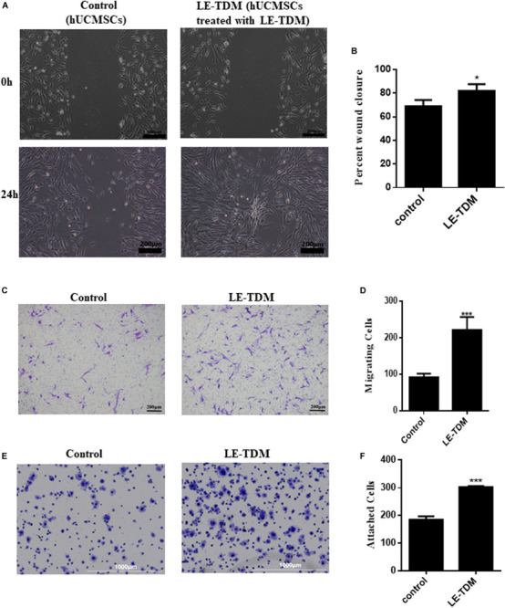
FIGURE 2.

Cell motility and adhesion ability of hUCMSCs treated with LE-TDM in comparison with hUCMSCs treated with regular media. A wound healing assay was performed, and the migration of the cells into the wound area was followed by microscopy at T = 24 h (A). Data are presented as the percentage of wound closure. Values are expressed as the mean ± SD (*P < 0.05) (B). Migrated hUCMSCs were visualized using crystal violet (C). The number of migrated cells was increased in the LE-TDM-treated group compared with that in the control group. Values are expressed as the mean ± SD (***P < 0.001) (D). LE-TDM affects hUCMSC adhesion. hUCMSCs attached to Fn-coated plates were visualized using crystal violet (E). The number of cells attached to Fn-coated plates was counted. Values are expressed as the mean ± SD (***P < 0.001) (F).
