Fig. 3.
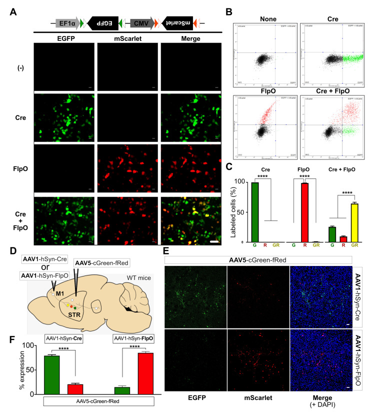
Validation of dual color gene expression vector in vitro. (A) Representative images showing Cre- or FlpO-selective expression of EGFP or mScarlet from the dual FP-expression vector. HEK293T cells were transfected with AAV-cGreen-fRed and pAAV-hSyn-Cre, and/or pAAV-hSyn-FlpO. EGFP or mScarlet expression was tested 72 h after transfection; scale bar: 50 μm. (B) Representative fluorescence-activated cell sorting (FACS) analysis showing recombinase-specific expression of EGFP or mScarlet in HEK293T cells co-transfected with the Cre and/or FlpO constructs. FACS-sorted cells with EGFP or mScarlet expression are displayed as colored scatter dots. (C) Bar graphs depicting the summary of FACS analysis results, showing the average percentage of EGFP (G), mScarlet (R), or EGFP+mScarlet (GR) - expressing cells from total transfected cells (mean±SEM). ****p<0.0001, one-way ANOVA with Tukey’s multiple comparison test. (D) Experimental scheme for in vivo validation of recombinase-specific expression of EGFP or mScarlet from AAV5-cGreen-fRed. AAV1-hSyn-Cre or AAV1-hSyn-FlpO was injected into M1 individually, but AAV5-cGreen-fRed was injected into the dorsal striatum. (E) Representative images showing M1-derived Cre- or FlpO-selective expression of EGFP or mScarlet from AAV5-cGreen-fRed in the striatum; scale bar=50 µm. (F) Bar graphs depicting the average percentage of EGFP (green)- or mScarlet (red)-expressing cell numbers (% expression) in the AAV5-cGreen-fRed-injected striatum when AAV1-hSyn-Cre or AAV1-hSyn-FlpO was injected into M1. N=seven slices randomly sampled from three mice. Mean±SEM. ****p<0.0001, unpaired t-test.

