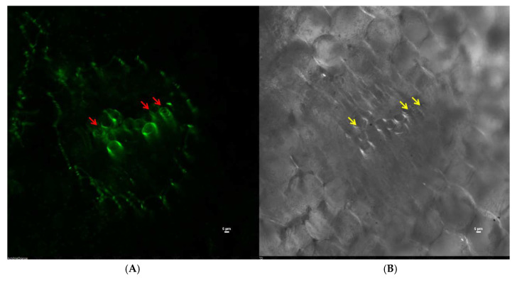
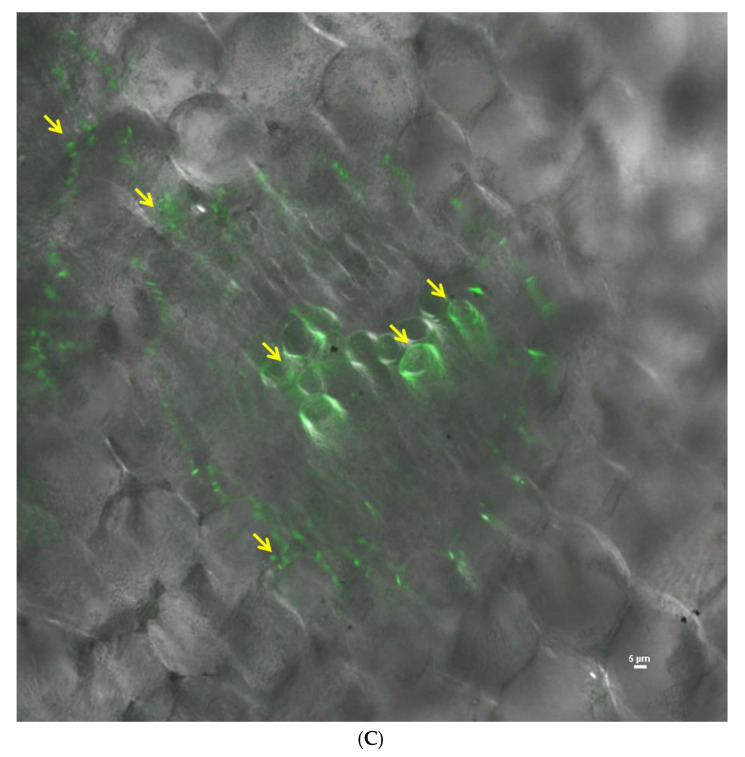
Figure 8.
Confocal scanning laser microscope images showing colonization of gfp-tagged R. solanacearum NAIMCC-B-01630 in tomato root; red arrows indicate vascular colonization of the tagged pathogen in 488 channel, and yellow arrows indicate the same in the TD channel (A) vascular colonization as seen in the 488 nm channel, (B) vascular colonization as seen in TD channel, and (C) superimposed image of 488 and TD channels.


