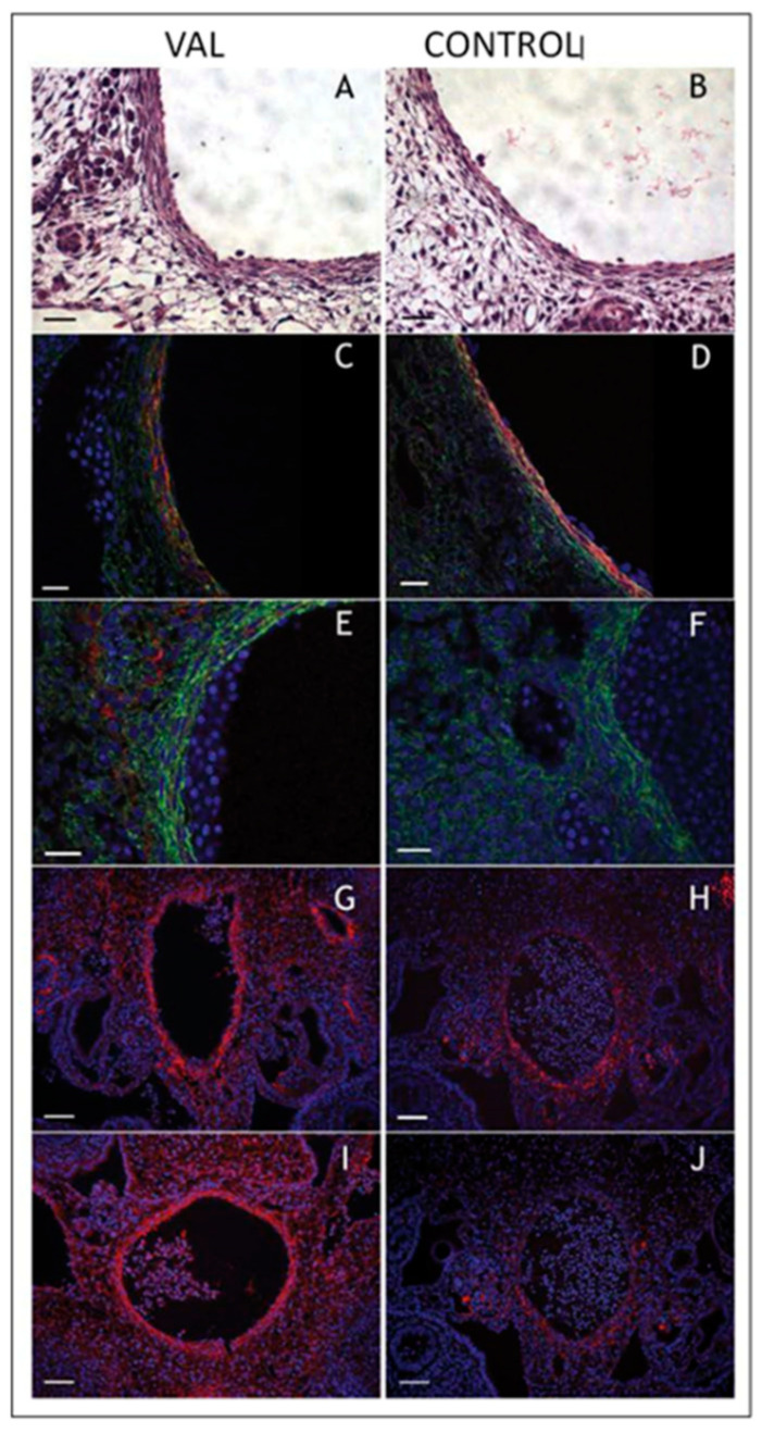
Figure 14.
Increased arterial load via unilateral vitelline artery ligation (VAL) alters aortic structural properties. Representative images of VAL (A,C,E,G,I) and control (B,D,F,H,J) dorsal aortas stained with hematoxylin and eosin (A,B), and antibodies against smooth muscle α-actin (C,D), collagen type III (E,F), procollagen type I (G,H), and antibody M38 (I,J) show increased content in dorsal aorta and perivascular tissues in VAL embryos. Magnification is × 600 and scale bars are 20 µm for A–F, and × 200 and 50 µm, respectively, for G–J. This was adapted with permission [221].

