
Figure 6.
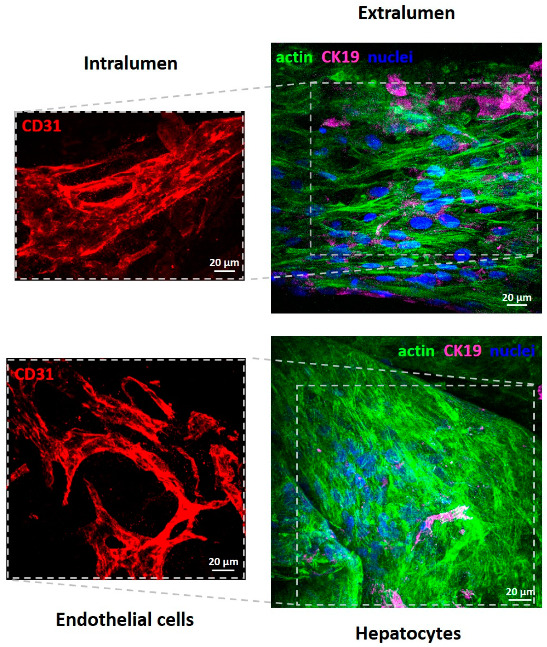
Confocal Laser Scanning Microscopy (CLSM) images of primary human hepatocytes and human endothelial cells after 18 days of culture in PCL HF membrane bioreactor. Endothelial cells in the intraluminal compartment were visualized for CD31 (red); hepatocytes over and between the fibers in the extraluminal compartment were stained for actin (green), CK19 (magenta), and nuclei (blue).