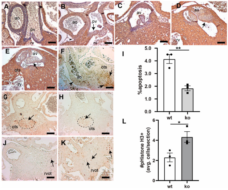
Figure 3.
Loss of Tgfb3 in mice results in the outflow tract cushion and vascular wall defects. (A–E), Cross-sections of E15.5–16.5 fetuses showing cardiac muscle actin (clone HHF35) immunohistochemistry for wildtype controls (A,C) and TGFβ3-deficient fetuses (B,D,E). Tgfb3−/− fetuses develop dysmorphic pulmonary (B) and aortic valve (D,E), and abnormal ascending aortic and pulmonary trunk walls (B) morphology. Tgfb3−/− fetuses also demonstrating hypoplastic outlet septum (E). (F) Representative image of a wildtype embryo (E11.5) using RNAscope in situ hybridization reveals Tgfb3 expression (brown punctate dots) in the vascular wall (arrowhead) and outflow tract septum (arrow). (G,H), Apoptosis (E13.5) using TUNEL staining (brown colored nuclei) in outflow tract septum mesenchyme. Compared to wildtype controls (G), Tgfb3−/− OFT septum have a reduced number of apoptotic cells (H). (I) Quantification of fraction of cells undergoing apoptosis. Mean ± SEM of % average apoptosis from at least 4 sections for each sample was used for comparison. Quantification was predominantly done in the area of outflow tract septum marked by a dotted circle. Reduced apoptosis in Tgfb3−/− hearts was noted as compared to wildtype embryos (** p = 0.004, Student’s t test; p = 0.07, Nonparametric (Mann Whitney test)). (J–L), Cell proliferation (E13.5) using phospho-histone H3 (Ser10) immunohistochemistry. Mean ± SEM of average pHistoneH3+ cells/section from at least 4 sections for each sample was used for comparison. Quantification was mainly restricted to the region around the fibrous outflow tract septum (L). Increased cell proliferation in Tgfb3−/− hearts (K, arrows) was observed as compared to wildtype (J) embryos (* p = 0.04, Student’s t test; p > 0.05, Nonparametric (Mann Whitney test)). Scale bars = 100 µm for (A–E,F–H,J,K). Abbreviations: rv, right ventricle; av, aortic valve; pv, pulmonary valve; ao, ascending aorta; ots, outlet septum, rvot, right ventricular OFT.

