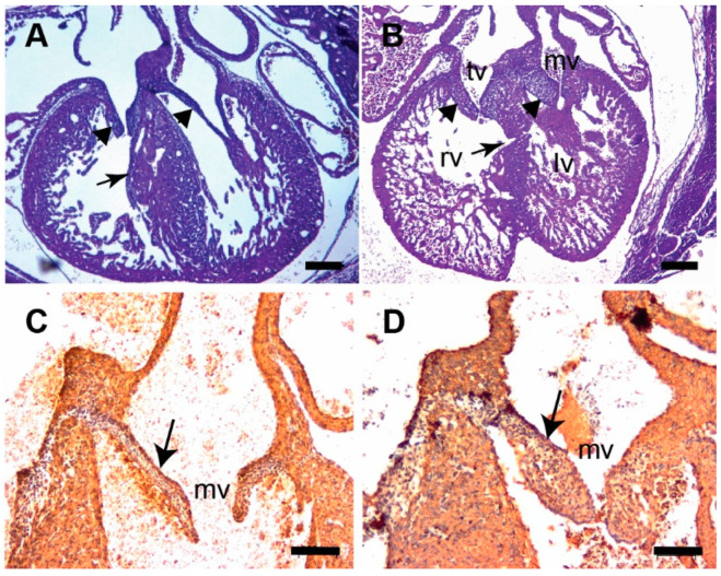
Figure 4.
Tgfb3 knockout fetuses exhibit atrioventricular valve thickening. (A,B) H&E stained sections of wildtype (A) and Tgfb3−/− fetus (E14.5-15.5) showing mitral valve and tricuspid valve thickening (B, arrowheads). Note that the ventricular myocardium is thin and abnormal with muscular ventricular septal defect in Tgfb3−/− (B, arrow). (C,D), Cardiac muscle actin (clone HHF35) immunohistochemistry of cross-sections of E15.5-16.5 wildtype (C) and Tgfb3−/− (D) fetuses. Tgfb3−/− fetus develop thickened mitral valves (D, arrow). Scale bars = 200 µm for (A,B), 50 µm for (C,D). Abbreviations: rv, right ventricle; lv, left ventricle; tv, tricuspid valve; mv, mitral valve.

