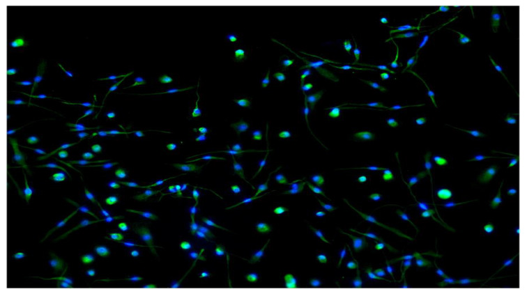
Figure 3.
The positive expression of CD14 by immune-fluorescence analysis, which is considered to be a typical marker for the identification of macrophages. After a 7-day culture, the cells were fixed with 4% paraformaldehyde for 15 min, permeabilized with PBS (1×) containing either 0.1% Triton X-100, labeled with anti-CD14 (green stain CD14 expression with Alexa Fluor 488) polyclonal antibody at a dilution of 1:50, followed by a goat anti rabbit Alexa Fluor® 488 secondary antibody to visualize the membrane. Images were assessed by inverted microscopy (Axio Observer Z1, Zeiss, resolution 100×). Nuclei were stained with DAPI. As a consequence of the growth factors used, GM-CSF led to a majority of elongated, fibroblast-spindle like shaped cells, and similar to macrophages existent in lung alveoli, in contrast the presence of M-CSF induced a majority of round or oval macrophages (fried eggs) like peritoneal macrophages.

