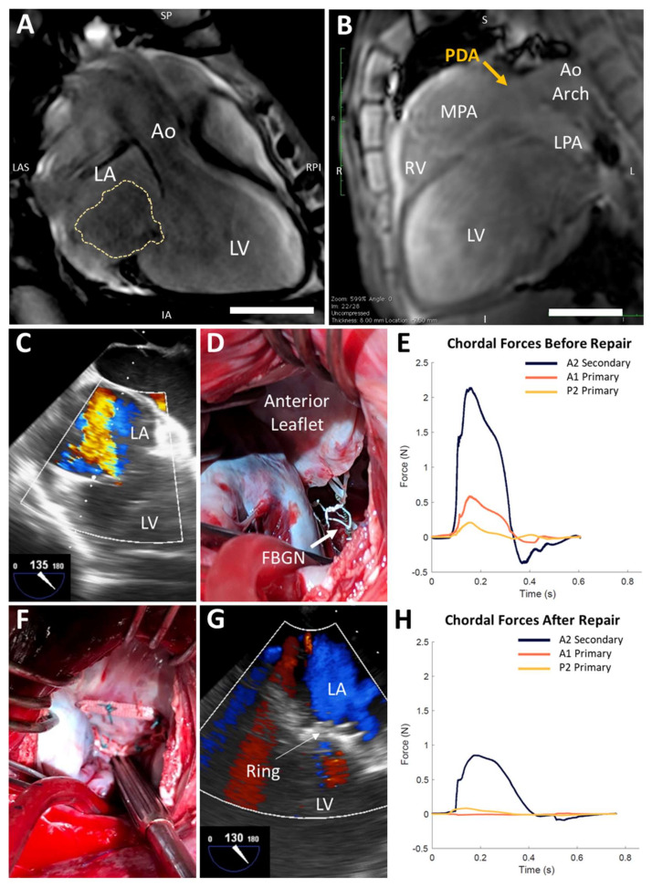
Figure 1.
Chordal forces are reduced after mitral ring annuloplasty in a rare ovine case of natural severe functional mitral regurgitation. (A,B) Cardiac magnetic resonance imaging reveals left atrial (LA) and left ventricular (LV) dilation, central mitral regurgitation (MR, yellow trace), and a patent ductus arteriosus (PDA). Aorta (Ao); inferior (I); inferior-anterior (IA); left (L); left anterior-superior (LAS); left pulmonary artery (LPA); main pulmonary artery (MPA); right (R); right posterior-inferior (RPI); right ventricle (RV); superior (S); superior-posterior (SP). Scale bars, 5 cm. (C) Baseline transesophageal echocardiography (TEE) confirms severe MR. (D) Instrumentation of native mitral valve chordae with force-sensing fiber Bragg grating neochordae (FBGN, white arrow). (E) Chordal forces in the state of severe MR over a representative cardiac cycle. (F) Mitral ring annuloplasty repair. (G) TEE confirms no MR after annuloplasty ring implantation. (H) Chordal forces are reduced after annuloplasty ring implantation.

