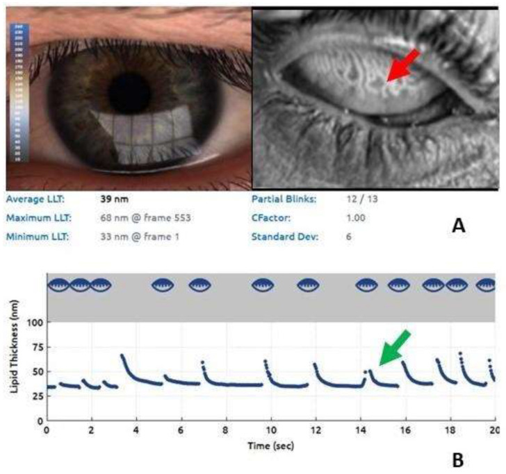
Figure 3.
Lipid imaging report and meibography using the Lip iView II (TearScience, Morrisville, NC, USA) ocular surface interferometer. (A) Remarkable alterations of the meibomian glands, the dilation of their bodies, the tortuosity of the glandular ducts, and the loss of glandular tissue (blue arrow). (B) Significant reduction of the LLT (39 nm) (red arrow).

