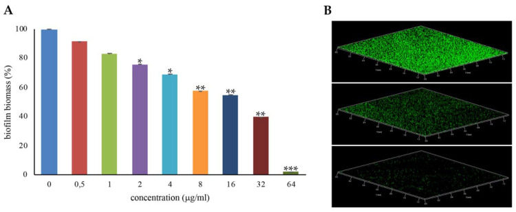
Figure 4.
Inhibition of S. aureus ATCC29213 biofilm formation with l-NPDNJ. (A) Biofilm was quantified after crystal violet staining. Values are presented as means ±SDs. Asterisks indicate statistically significant differences between treated and untreated biofilms (* p < 0.05, ** p < 0.01, *** p < 0.001). (B) Confocal laser scanning microscopy (CLSM) analysis of the biofilm formed in the absence (upper panel) or presence of l-NPDNJ at the concentrations of 32 (middle panel) and 64 μg/mL (inferior panel).

