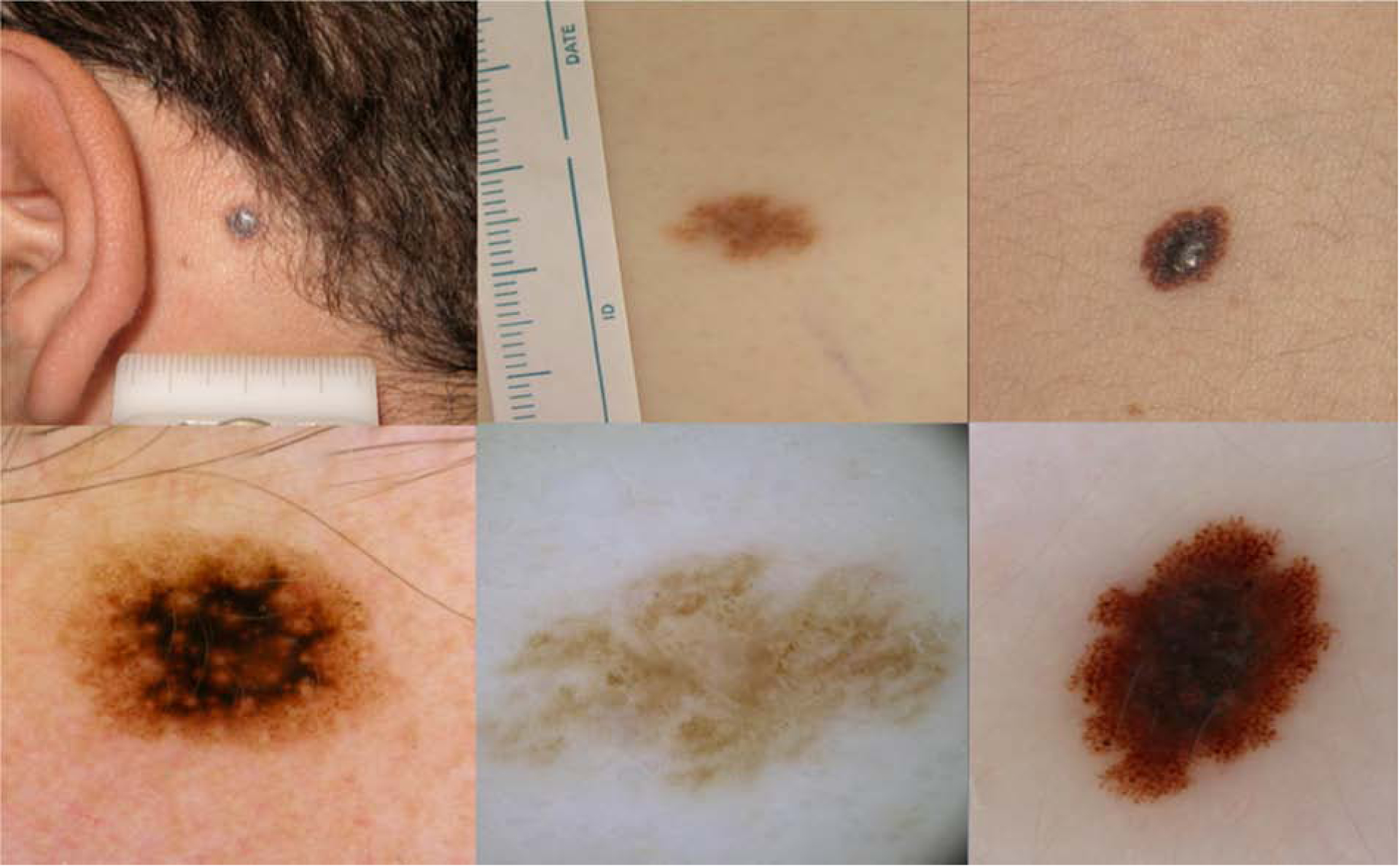
Figure 2. Pattern 2, Nevus-like. Non-Spitzoid melanomas in adolescents.

a) Clinically, symmetric 5mm lesion on a 19 year old boy.
b) Dermoscopy: Symmetric globular pattern, with structureless central black area. Superficial spreading Melanoma, Breslow 0.6mm.
c) Clinically, symmetric light brown macule on the chest of a 17year old boy affected by multiple atypical nevi and previous melanoma.
d) Dermoscopy: Symmetric light brown lesion showing irregular globules and negative network. Melanoma arising in a nevus, Breslow 1.0mm.
e) Clinically asymmetric lesion on the trunk of a 19 year old boy.
f) Dermoscopy: symmetric reticular-globular pattern, with atypical network, irregular pigmented globules and dots at the periphery and central blue-white veil. Melanoma arising in a nevus, Breslow 3.1mm.
