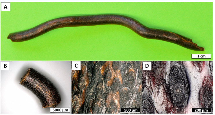
Figure 1.
Overview of the Cirrhipathes sp. coral fragments used in the study. (A) Central portion of the unbranched, unpinnulated stem of the colony. (B) Close-up view of the skeletal surface showing the multiple longitudinal rows of spines. (C) Basal plates of the spines after erosion. (D) Close-up view of one spine basal plate showing the concentric layers of skeleton.

