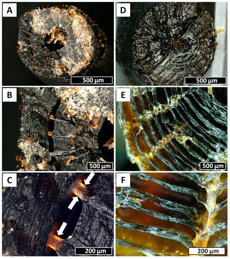
Figure 2.
Insights into the inner structure of Cirrhipathes sp. skeleton. (A) Transversal section of the stem, showing a clearly hollow central canal surrounded by concentric layers of skeleton. The outer surface is covered in small triangular spines. (B,C) Spines’ roots visible between the skeletal concentric layers. (D) Transversal section of the stem with a central canal partially closed by a skeletal septum. (E,F) Clusters of concentric skeletal layers intersecting perpendicularly with the spines’ roots, connecting vertically the outer surface with the internal channel.

