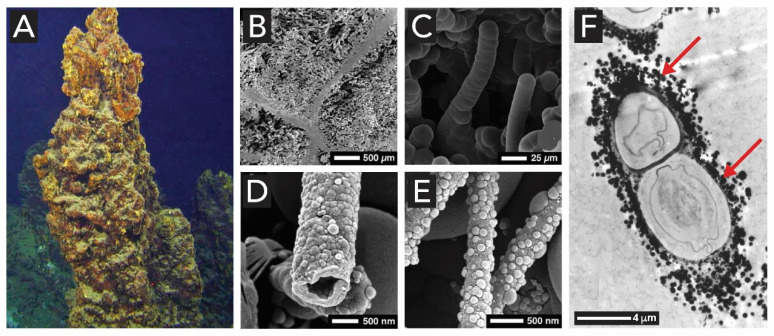
Figure 7.
Silicified microbes. (A) Underwater photographs of silica-rich chimneys (~2 m) on the Giggenbach volcano. (B–E) SEM photomicrographs of silicified microbes: (B) Overview of silicified microbial mats with opal-A sheets. (C) Silicified filamentous microbe. (D) Silicified rod-shaped microbe showing a thin wall around an open lumen and small opal-A spheres on the outer surface. (E) Silicified rod-shaped microbes coated with opal-A and scattered opal-A spheres. (F) TEM cut of silicified bacteria from a geyser outflow channel, showing a filamentous cyanobacterium with silica spheroids on outer sheath (arrows). Images A–E are adapted from [79] with permission from John Wiley and Sons. Image F is reproduced from [80] with permission from Springer Nature BV.

