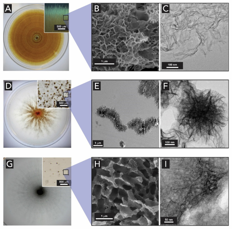
Figure 25.
Optical microscopy, SEM, and HR-TEM images of Mn oxides produced by Plectosphaerella cucumerina DS2psM2a2 (A–C), Stagonospora sp. SRC1lsM3a (D–F) and Acremonium strictum DS1bioAY4a (G–I) growing radially outward from the inoculation point in the center of the petri dishes. (A,D,G) Optical micrographs of thin sheets of Mn oxides on their surface. (B,E,H) SEM images of the Mn oxides particles produced by the fungi. (C,F,I) HR-TEM images showing the morphology of Mn oxides. Adapted from [185] with permission from Elsevier.

