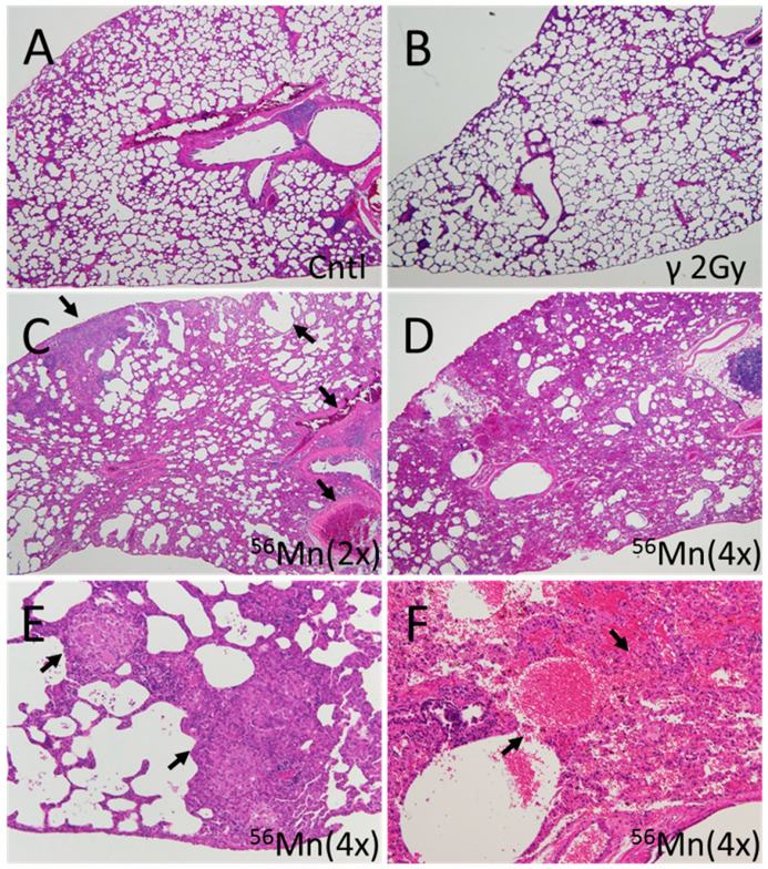
Figure 3.
Representative fields of lung tissue late histology from 56Mn exposed, 2.0 Gy γ exposed and Control rats collected at month 6 post-irradiation. (A) Lung of a control rat. No pathologic changes were observed; (B) Lung of a 2.0 Gy γ exposed rat. No pathologic changes were observed; (C) Lung of a 56Mn(2×) exposed rat. Widespread emphysema and atelectasis as well as hemorrhage with vessel thrombosis were observed (arrows); (D) Lung of a 56Mn(4×) exposed rat. More extensive damage, pneumonitis resulting from severe inflammation and inflammatory cell infiltration as well as intra-alveolar hemorrhage were observed; (E) Lung of a 56Mn(4×) exposed rat. Arrows indicate granuloma surrounded by emphysema was observed in a lung lobe; (F) Lung of a 56Mn(4×) exposed rat. Arrows indicate severe hemorrhage and inflammatory cell infiltration. A higher magnification of D. H&E stain, 4× (A–D) and 20× (E,F) objective magnification.

