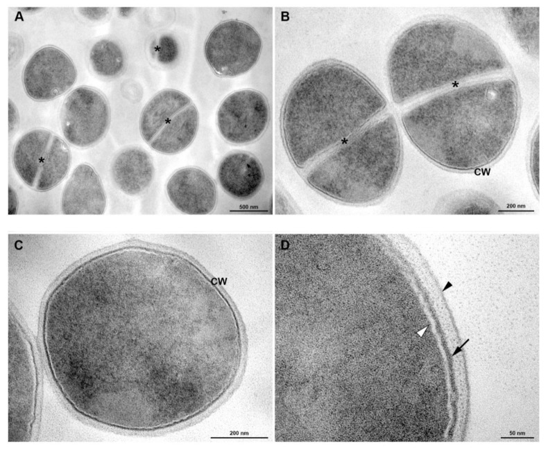
Figure 5.
Transmission electron microscopy of S. aureus grown under culture medium. (A) General view showing rounded cells with a thick cell wall envelope and homogeneous electron density in the cytoplasm. Central division septa (*) are seen in some cells. (B,C) Detailed view of non-dividing (B) and dividing (C) bacteria A tripartite cell wall (CW) is seen enclosing the plasma membrane. Asterisk indicates the septum. (D) Inset of the cell wall. Black arrowhead indicates outer highly stained fibrous surface and intermediate translucent region; Arrow points to a heavily stained inner thin zone; the plasma membrane (white arrowhead) is seen immediately below this electrodense layer of the wall. Bars: A: 500 nm; B,C: 200 nm; D: 100 nm.

