Figure 8.
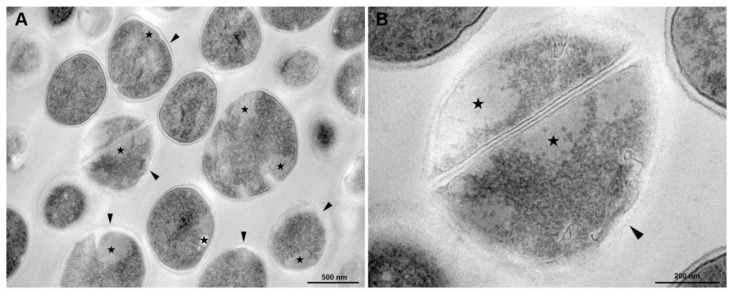
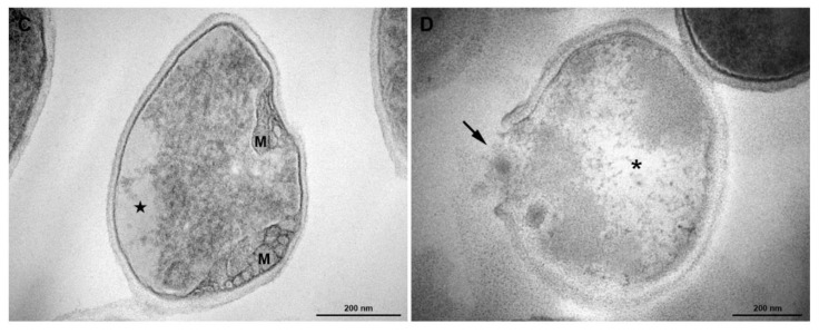
Transmission electron microscopy of S. aureus treated with antibiotics (Menadione MIC/4) and Antibiotic CIM/4). (A) General view: some cells exhibit abnormal electron density in the cytoplasm (★) and altered cell wall with no visible tripartite-layers structure (black arrowheads). (B) Inset of Figure A. (C) Detailed view of a cell showing alterations in the shape, loss of cytosolic electron-density (★) and mesosome-like structures (M). (D) A lysed cell with cell wall disruption (arrow) and cytoplasmic disintegration (asterisk). Bars: A: 500 nm; B–D: 200 nm.


