Figure 1.
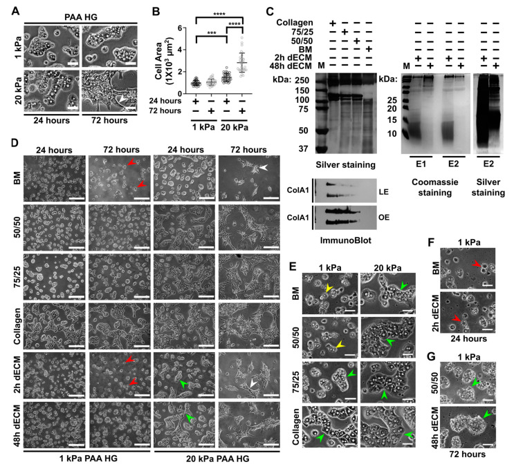
Collagen type I is necessary for cell adhesion and aggregation in rat primary hepatocytes cultured on soft polyacrylamide hydrogels (PAA HGs). (A) Magnifications of representative differential interference contrast (DIC) images of primary hepatocytes cultured on soft (1 kPa) and stiff (20 kPa) polyacrylamide hydrogels (PAA HGs) conjugated with collagen type I after 24 and 72 h of culture. Scale bar: 50 μm. (B) Quantitative analysis and comparison of cell area of hepatocytes cultured on soft and stiff PAA HGs at 24 h and 72 h of culture. Data are represented as mean ± standard deviation (SD) of a representative of 3 independent experiments, **** p < 0.0001, *** p < 0.001 (C) Silver staining of SDS-PAGE of Matrigel basement membrane-associated matrix (BM) and collagen type I at different proportions (left) and decellularized liver matrices (dECM) extracted after 2 h and 48 h of digestion (right). Lower panel: collagen type I immunoblotting in collagen and BM mixtures (below). Membranes low-exposed (LE) and over-exposed (OE) (lower panel). In addition, Coomassie staining of 2 h and 48 h dECM of 2 independent experiments (E1 and E2) ran in parallel. (D) Primary hepatocytes cultured on 1 kPa and 20 kPa PAA HGs conjugated with different collagen I/BM proportions and decellularized matrix extracts (dECM) at 24 h and 72 h of culture. Images were obtained from a 20× objective. Scale bar: 200 μm. (E) Magnifications of differential interference contrast (DIC) images of primary hepatocytes cultured on soft and stiff PAA HGs at 72 h of culture. Collagen and BM were conjugated at different proportions. Scale bar: 50 μm. (F–G) Magnifications of DIC images of hepatocytes cultured on soft PAA HGs conjugated with different extracellular matrices (ECMs) at 72 h of culture. ECM mixtures: BM, 100% matrigel; 50/50, 50% collagen type I plus 50% BM; 75/25, 75% collagen type I plus 25% BM; collagen, 100% collagen type I; 2h dECM, dECM after 2 h of digestion; 48h dECM, ECM after 48 h of digestion; M, molecular weights. Scale bar: 50 μm. Aggregates of primary hepatocytes: green arrowheads; apoptotic bodies: red arrowheads; individual hepatocytes: yellow arrowheads; transformed hepatocytes: white arrowheads.

