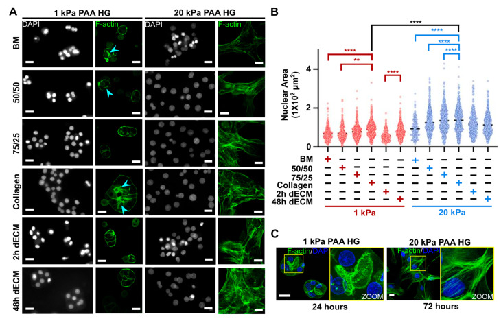
Figure 2.
Actin cytoskeleton remodeling and nuclear area increase are inhibited in primary hepatocytes cultured on soft PAA HGs. (A) Representative fluorescence images of nuclei (4′,6-diamidino-2-phenylindole, or DAPI) and actin filaments (F-actin) in hepatocytes after 72 h of culture on soft and stiff PAA HGs conjugated with ECM matrices. Actin filaments proximal to canaliculi-like structures: light blue arrowhead. Images were obtained from 40× (epifluorescence) and 63× (confocal microscopy) objectives for nuclei and F-actin, respectively. Scale: 20 µm. (B) Quantitative analysis of nuclear area in primary hepatocytes cultured on soft and stiff PAA HGs linked to different ECM. ECM mixtures: BM, 100% Matrigel; 50/50, 50% collagen type I plus 50% BM; 75/25, 75% collagen type I plus 25% BM; collagen, 100% collagen type I; 2 h dECM, dECM after 2 h of digestion; 48 h dECM: ECM after 48 h of digestion. Significant differences are shown in red and blue between PAA HG with the same elastic condition; black lines show significant differences between PAA HGs with the different elastic conditions. Data are represented as mean ± SD of a representative of two independent experiments, ** p < 0.01, **** p < 0.0001. (C) 3D projections of confocal images of hepatocytes cultured on 1 kPa and 20 kPa PAA HGs at 24 h and 72 h after culture, respectively. Canaliculi-like structures and stress fibers are highlighted and magnified at yellow squares (right panels). Scale bars: 20 µm.

