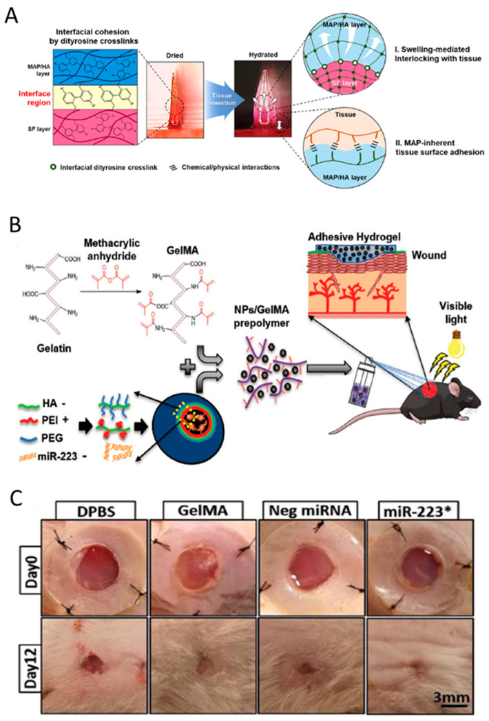
Figure 6.
(A) Illustration of the mechanisms of a hydrogel-forming adhesive microneedle patch consisting of a mussel adhesive protein-based swellable and sticky shell and a silk fibroin-based non-swellable core. It shows the structure and the reaction of the fabricated patches at the interface with the tissue. Adapted with permission from Jeon et al. [143]. (B) Synthesis illustration of the process for the formation of hyaluronic acid nanoparticle/miR-223*-laden GelMA hydrogels and the use of these adhesive hydrogels for wound healing. Adapted with permission from Gao et al. [144]. (C) Images of the wounds treated with Dulbecco’s phosphate-buffered saline (DPBS), GelMA, Neg miRNA incorporated GelMA, and miR-223* incorporated GelMAat days 0 and 12 of treatment. * Images for animals at day 12 were acquired after removal of the silicon splint to highlight the extent of wound healing. Adapted with permission from Saleh et al. [144].

