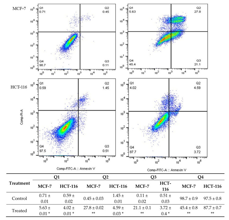
Figure 2.
The level of apoptosis and necrosis was assessed by flow cytometry using Annexin V/PI staining, the control groups were without any treatment, and the M. fruticosa extract groups were treated with 100 μg/mL M. fruticosa extract for 48 h, * p < 0.05, ** p < 0.001. Q1: necrotic cells, Q2, late apoptotic, Q3: early apoptotic, Q4: living cells.

