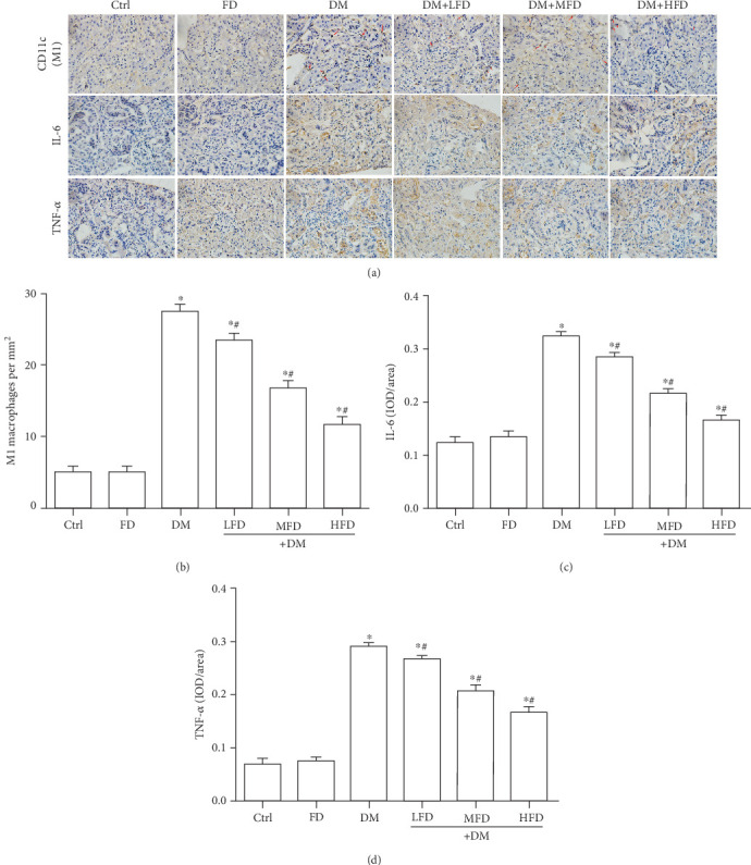
Figure 3.

Fasudil inhibited M1 macrophage polarization and proinflammatory cytokines in renal tissues of diabetic mice. (a) Representative immunohistochemical staining of CD11c (a marker for M1 macrophages; red arrows), IL-6, and TNF-α in renal tissues. (b) Quantitative analysis for the number of M1 macrophages in renal tissue specimens. Data are expressed as mean ± SD, ∗P < 0.05 vs. the Ctrl group; #P < 0.05 vs. the DM group. (c) Quantitative analysis of the protein expression of IL-6 in renal tissue specimens. Data are expressed as mean ± SD, ∗P < 0.05 vs. the Ctrl group; #P < 0.05 vs. DM group. (d) Quantitative analysis of the protein expression of TNF-α in renal tissue specimens. Data are expressed as mean ± SD, ∗P < 0.05 vs. the Ctrl group; #P < 0.05 vs. the DM group.
