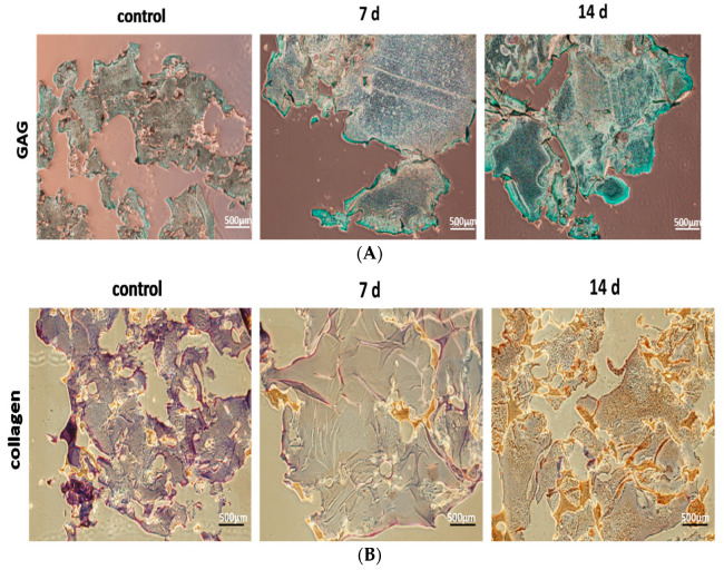
Figure 6.
Immuno-histochemical (IHC) analysis of hBMSCs in the model hydrogels after hBMSC were induced by TGF- to differentiate them to NP cells for 7 d and 14 d, respectively. Deposition of (A) GAG (in blue) and, (B) Collagen type II (in brown) in the model hydrogels were shown which were significantly higher than those of cultivated in cultural wells (e.g., control group).

