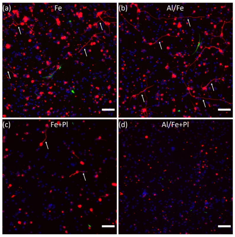
Figure 4.
Representative fluorescence images of β-tubulin III labelled (red) neurons and neurites (arrows), GFAP labeled glia (green), and cell nuclei (blue) on all VACNT preparations. (a) Fe, (b) Al/Fe, (c) Fe + Pl, and (d) Al/Fe+Pl showing the occurrence of neurite-bearing cells and the extent of neurite outgrowth in the different preparations. Scale bars: 50 µm.

