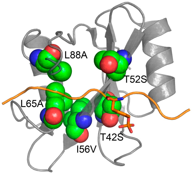
Figure 2.
Three dimensional structure of the N-SH2 domain of SHP2 (in gray) in complex with Gab1 (in orange (Gab1 sequence: NTERM-GDKQVEYLDLDLD-CTERM [PDB:4QSY])). Residues T42, T52, I56, L65 and L88 of N-SH2 are shown in green to highlight their physical location in respect to the binding pocket of the domain.

