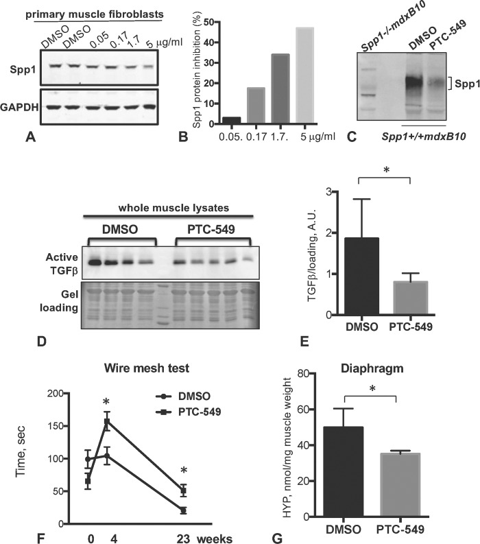
Figure 6.

Effect of postnatal pharmacological inhibition of Spp1 in mdxB10 mice. (A) Western blot of Spp1 levels in mdxB10 primary fibroblasts that were incubated with increasing concentration of PTC-549 for 72 h. Blot was probed with anti-Spp1 antibodies (upper panel). Lower panel shows GAPDH, which was used as a loading control. (B) Quantitative analysis of the western blot shown in (A). Bars represent the percent of Spp1 inhibition relative to DMSO control. (C) Western blot of whole muscle lysates from DMSO- or PTC-549-treated mice (n = 5 per group in pooled samples) probed with anti-Spp1 antibody. First lane shows muscle lysate lacking Spp1 to ensure antibody specificity. (D) Western blot analysis of total protein lysates from mdxB10 mice treated with PTC-549 or DMSO for 6 months (n = 4 per group). The data showed decreases in the active form of TGFβ (25 kDa) in PTC-54-treated mice. Quantitative analysis of the western blot is shown in (E). (F) Wire mesh test showed that time on the wire was significantly higher for mice treated with PTC-549 at 4 weeks of treatment and up to 23 weeks (n = 8 and 9 mice per group). (G) Analysis of total collagen content after 24 weeks of treatment with PTC-549 or DMSO using hydroxyproline (HYP) assay. Asterisk indicates statistical significance of P < 0.05; AU, arbitrary units.
