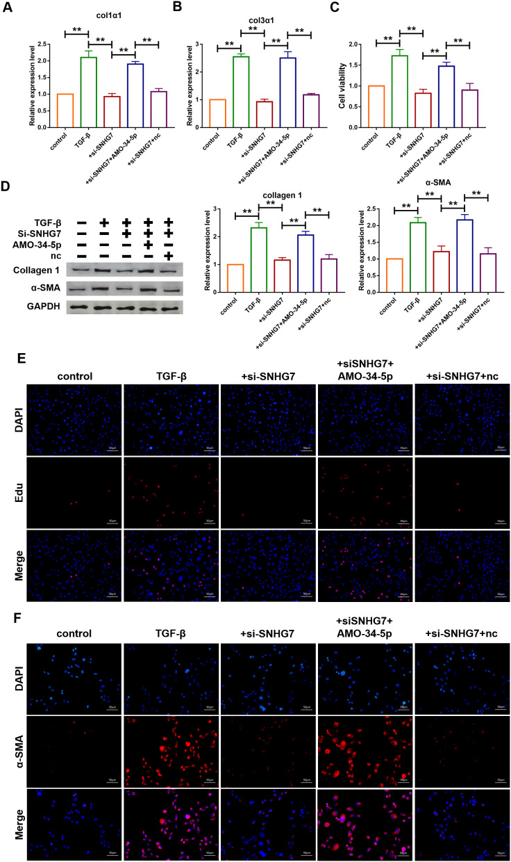
Figure 5.
Silencing of lncRNA SNHG7 alleviated TGF-β1-induced fibrogenesis in cardiac fibroblasts. (A, B) Suppression of SNHG7 attenuated the increase in collagen 1α1 and collagen 3α1 expression induced by TGF-β1, as measured by qRT-PCR; GAPDH mRNA served as an internal control. Data was presented as mean ± SEM; one-way ANOVA was used for the statistical analysis. n=5 independent cell cultures. (C) Western blotting analysis showing that knockdown of SNHG7 attenuated TGF-β1-induced fibrotic protein expression (Collagen I and α-SMA); GAPDH served as a loading control. Data was presented as mean ± SEM; one-way ANOVA was used for the statistical analysis. n=5 independent cell cultures. (D) MTT assay for the assessment of cell viability. Transfection of si-SNHG7 with or without AMO-34-5p in cardiac fibroblasts treated with TGF-β1 for 24h. Data was presented as mean ± SEM; one-way ANOVA was used for the statistical analysis. n=5 independent cell cultures. (E) EdU staining for the assessment of cell proliferation in cardiac fibroblasts inhibiting SNHG7 in the presence or absence of AMO-34-5p mimics. Scale bars represented 50 μm. (F) Representative images of immunofluorescence staining showing that knockdown of SNHG7 abated the TGF-β1-induced fibroblast-myofibroblast transition, which was promoted by AMO-34-5p. Scale bars represented 50 μm. **P<0.05.

