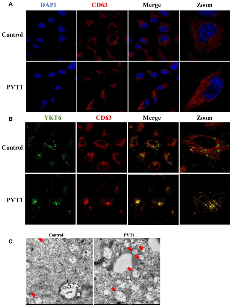
Figure 3.
PVT1 promotes the movement of MVBs towards the plasma membrane. (A) Analysis of CD63 (red) in PVT1-overexpressing HS766T cells, as determined by confocal microscope. Nuclei were labeled with DAPI (blue). (B) Analysis of YKT6 (green) and CD63 (red) in PVT1-overexpressing HS766T cells, as determined by confocal microscope. (C) Exosomes in PVT1-overexpressing HS766T cells, as determined by electron microscope.

