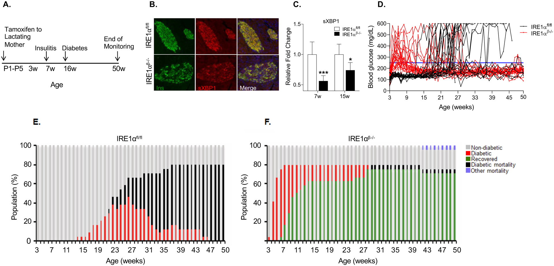
Figure 1. IRE1αβ−/− NOD female mice are protected from T1D.

(A) Schematic representation of tamoxifen-induced deletion of IRE1α in β-cells of NOD mice.
(B) Representative immunofluorescence images showing sXBP1 expression on pancreatic sections from 5-week-old mice.
(C) Quantification of sXBP1 expression in the islets of 7- and 15-week-old IRE1αfl/fl (7 weeks: n = 6; 15 weeks: n = 5) and IRE1αβ−/− mice (7 weeks: n = 5; 15 weeks: n = 6). Data are averages of two technical replicates from a representative experiment.
(D) Blood glucose levels of IRE1αfl/fl and IRE1αβ−/− mice (n = 24 per group).
(E and F) Diabetes progression in IRE1αfl/fl and IRE1αβ−/− mice.
All data are represented as mean ± SEM, with statistical analysis performed by Student’s t-test (***P < 0.001, *P < 0.05).
