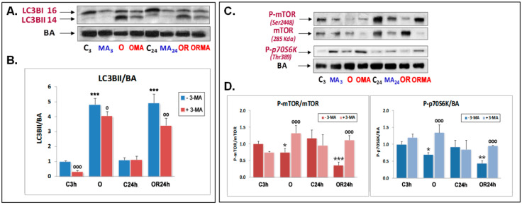
Figure 1.
Effect of oxygen and glucose deprivation (OGD), oxygen and glucose deprivation and resupply (OGDR), and 5 mM 3-methyladenine (3-MA) treatment on LC3B lipidation (A,B) and mTOR and p70S6K phosphorylation (C,D) in cultured cortical neurons. (A,B) Western blot analysis of LC3BI and LC3BII was performed in extracts of cortical neurons subjected to 3 h OGD exposure (O) and subsequent 24 h OGDR (OR) compared to their respective controls. (A) Representative LC3B I and II and β-actin (BA) Western blots are shown. (B) Quantitative analysis of the LC3BII/BA ratio. Data are expressed as ratios over basal control and are mean ± SEM of three experiments, each one performed in duplicate. (C,D) Western blots of mTOR, 2448SerP-mTOR, and 389ThrP-p70S6K were performed in the aforementioned conditions. Upper panel (C) shows a representative Western blot of these proteins. Lower panels (D) shows the quantification of the indicated ratios and are mean ± SEM of three experiments, each one performed in duplicate. Statistics in panels (B) and (D) compare the effect of O and OR over their respective controls C3 and C24h, respectively (***) and the effect of 5 mM 3-MA in each condition, in the absence or presence of this compound (ooo), at */o p < 0.05, **/oo p < 0.01, and ***/ooo p < 0.001) (one-way ANOVA test).

