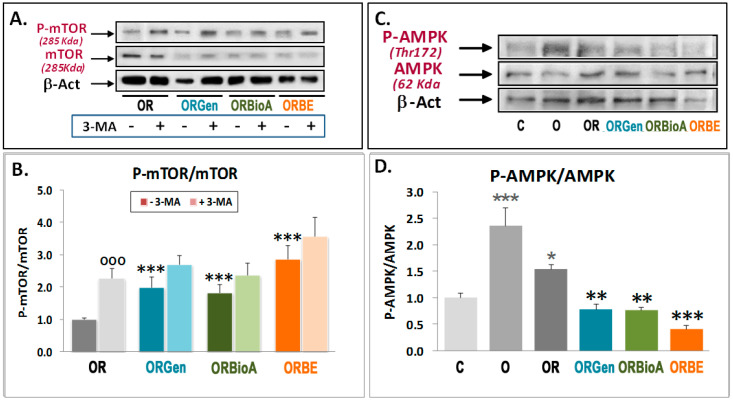
Figure 4.
Effects of OGDR, phytoestrogens, and 3-MA treatment on mTOR and AMPK phosphorylation. Western blots of mTOR, 2448SerP-mTOR (A,B), and 172Thr-P-AMPK (C,D) were performed in extracts of cortical neurons exposed to 3 h OGD exposure (O) and 24 h OGDR (OR) in the absence or presence of 5 mM 3-MA (A,B) or under OR conditions in the absence or presence of 1 μM Genistein (Gen), BiocaninA (BioA), and 17-β-estradiol (BE)). Upper panels (A,C) show representative Western blots. Lower panels (B,D) represent the quantitative analysis of indicated ratios and are mean ± SEM of three experiments, each one performed in duplicate. Statistics comparing the effect of 5 mM 3-MA in each condition versus respective conditions in the absence of this compound (ooo) panel (B), the effect of O and OR over control C3 (***) panel (D), and the effect of phytoestrogens on OR condition without them (***) panels (B,D), at * p < 0.05, ** p < 0.01, and *** p < 0.001 (one-way ANOVA test) are shown.

